

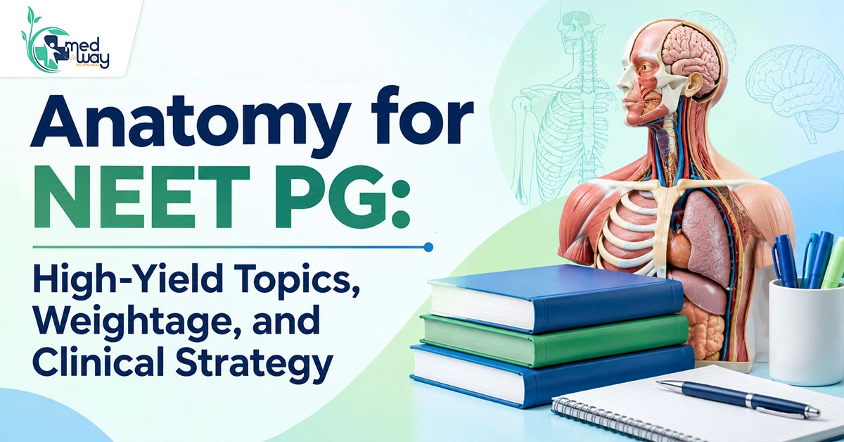
Anatomy for NEET PG 2026: High-Yield Topics, Weightage, PYQs, Books & 30-Day Preparation Strategy
Anatomy for NEET PG 2026 is much more than a subject of muscles, nerves, vessels and anatomical relations. It forms the foundation for understanding several clinical subjects, including Surgery, Orthopaedics, Neurology, Radiology, ENT, Ophthalmology, Obstetrics and Gynaecology, and Pediatrics. A strong understanding of Anatomy can therefore help you solve not only direct Anatomy questions but also clinically integrated questions across the NEET PG paper.
For example, a question about a fracture of the surgical neck of the humerus may require you to identify the axillary nerve, its anatomical course, the muscles it supplies and the resulting sensory deficit. Similarly, questions on inguinal hernia, thyroid surgery, brainstem stroke syndromes or pelvic surgery often require you to apply anatomical relationships rather than simply recall an isolated fact.
The preparation strategy for Anatomy in NEET PG has consequently shifted from extensive rote memorisation towards conceptual understanding, clinical correlation, image-based learning, PYQs, MCQs and repeated revision. Instead of trying to memorise every anatomical detail, aspirants should prioritise frequently tested concepts and understand how structures relate to one another in clinical situations.
Quick Overview: Anatomy for NEET PG 2026
| Particular | Details |
| Subject | Anatomy |
| Exam | NEET PG 2026 |
| Conducting body | National Board of Examinations in Medical Sciences (NBEMS) |
| Main preparation areas | Gross Anatomy, Neuroanatomy, Embryology, Histology, Genetics, Radiological and Clinical Anatomy |
| Best approach | Concept building + PYQs + MCQs + image practice + revision |
| High-priority areas | Neuroanatomy, brachial plexus, cranial nerves, embryology, abdomen, pelvis, thorax, head and neck |
| Important question formats | Clinical vignettes, image-based questions, anatomical relations and applied Anatomy |
NEET PG Anatomy Syllabus 2026
The NEET PG Anatomy syllabus should be studied as a combination of basic, regional, developmental, microscopic and applied Anatomy. The official NBEMS NEET-PG portal should be used for the latest examination bulletin and notices.
For preparation purposes, divide the subject into the following areas.
| Anatomy Area | Topics to Cover | Clinical Integration |
| General Anatomy | Anatomical terminology, tissues, bones, joints, muscles, vessels and nerves | Basic clinical understanding |
| Gross/Regional Anatomy | Upper limb, lower limb, thorax, abdomen, pelvis, head and neck | Surgery, Orthopaedics, ENT |
| Neuroanatomy | Brain, brainstem, spinal cord, cranial nerves, tracts and blood supply | Neurology, Neurosurgery |
| Embryology | Pharyngeal arches, heart development, gut rotation, organ development | Pediatrics, Surgery |
| Histology | Epithelium, connective tissue, cartilage and organ histology | Pathology |
| Human Genetics | Chromosomal abnormalities and clinically relevant syndromes | Pediatrics, Medicine |
| Radiological Anatomy | X-ray, CT, MRI and cross-sectional anatomy | Radiology, Surgery |
| Surface and Sectional Anatomy | Surface landmarks and anatomical structures in sections | Clinical examination and imaging |
NEET PG Anatomy Weightage 2026
There is no fixed official NBEMS document that guarantees a particular number of questions from individual Anatomy subtopics such as Neuroanatomy, Embryology or Gross Anatomy. Question distribution can change between examinations.
NEET PG Anatomy Topic Priority
| Topic | Priority | Important Areas |
| Neuroanatomy | Very High | Brainstem, cranial nerves, tracts, visual pathway, cerebral blood supply |
| Clinical/Gross Anatomy | Very High | Brachial plexus, nerve injuries, hernias, abdomen, pelvis, thorax |
| Embryology | High | Pharyngeal arches, heart development, gut rotation and congenital anomalies |
| Head & Neck | High | Neck triangles, thyroid, laryngeal nerves, cavernous sinus |
| Radiological Anatomy | High | CT, MRI, X-ray and cross-sectional anatomy |
| Thorax | High | Heart, coronary arteries, lungs, mediastinum and diaphragm |
| Abdomen & Pelvis | High | Vessels, peritoneum, inguinal canal and pelvic relations |
| Histology | Moderate–High | Epithelium, cartilage and organ identification |
| Genetics | Moderate | Chromosomal abnormalities and syndromes |
Most Important Anatomy Topics for NEET PG 2026
If you are searching for the most important topics in Anatomy for NEET PG, prioritise concepts that are repeatedly useful in clinical questions.
| No. | High-Yield Anatomy Topic | Important Areas to Focus On |
| 1 | Neuroanatomy |
|
| 2 | Brachial Plexus |
|
| 3 | Cranial Nerves |
|
| 4 | Embryology |
|
| 5 | Abdomen |
|
| 6 | Pelvis |
|
| 7 | Thorax |
|
| 8 | Head and Neck |
|
NEET PG Anatomy PYQs and Previous-Year Question Analysis
Anatomy PYQs for NEET PG are one of the most useful resources for understanding how concepts are tested.
Do not use PYQs only as a question bank. Use them to identify patterns.
For every PYQ, ask:
- What concept is being tested?
- Is it a factual or clinical question?
- Can the same concept be tested through an image?
- Which other subject is connected with it?
- Did I get it wrong because I lacked knowledge or because I misread the clinical clue?
Frequently Tested Anatomy Concepts
Give special attention to recurring themes such as:
- Brachial plexus
- Cranial nerves
- Brainstem
- Cerebral blood supply
- Spinal cord tracts
- Hernias
- Pelvic anatomy
- Embryology
- Nerve injuries
- Radiological anatomy
Clinical-Vignette Questions
Modern medical entrance questions may present Anatomy through a clinical situation.
For example:
A patient develops wrist drop following an injury to the arm.
Instead of asking directly about the radial nerve, the question may ask about:
- The injured nerve
- Its anatomical course
- The affected muscle
- The site of injury
- The associated sensory deficit
Anatomy MCQs for NEET PG: How to Practise
MCQs should come after concept building rather than replacing it.
A useful sequence is:
Read → Recall → Solve MCQs → Analyse mistakes → Revise
When solving Anatomy MCQs for NEET PG, maintain an error notebook containing:
- Incorrect questions
- Confusing anatomical relations
- Repeated facts
- Image-based mistakes
- PYQ concepts
Anatomy Image-Based Questions for NEET PG
Image-based questions have become an important part of modern medical entrance preparation. Candidates should be comfortable recognising structures in different imaging planes and clinical settings.
| No. | Topic | Important Areas to Practise/Revise |
| 1 | CT Anatomy |
|
| 2 | MRI Anatomy |
|
| 3 | X-Ray Anatomy |
|
| 4 | Cadaveric Anatomy |
|
| 5 | Histology Images |
|
| 6 | Cross-Sectional Anatomy |
|
Best Books for Anatomy for NEET PG
Choosing too many resources can actually reduce revision efficiency. Your goal should be to have one primary concept source, one revision source and one question source.
| Resource | Best Use |
| BD Chaurasia | Detailed gross anatomy and concept clarification |
| Gray’s Anatomy | Reference for difficult concepts |
| Revision Notes | Fast revision |
| PYQ Books | Previous-year question practice |
| MCQ/QBank | Clinical application and exam practice |
| Image Resources | CT, MRI, X-ray and cadaveric practice |
| 3D Anatomy Tools | Spatial relationships |
BD Chaurasia for NEET PG
BD Chaurasia can be useful for building concepts, especially in Gross Anatomy. However, reading every detail repeatedly may not be the most efficient strategy during the final stages of NEET PG preparation.
Use it selectively when you need to understand:
- Anatomical relations
- Nerve courses
- Vascular anatomy
- Regional anatomy
- Surgical relationships
Gray’s Anatomy
Gray’s Anatomy is an excellent reference but should not normally be your primary revision book for NEET PG.
Use it when a difficult anatomical relationship requires clarification.
Anatomy Notes for NEET PG
Your final Anatomy notes for NEET PG should be concise enough to revise repeatedly.
Include:
- Diagrams
- Tables
- Nerve supply
- Blood supply
- Embryology derivatives
- Clinical correlations
- Important exceptions
- Frequently incorrect PYQs
Best Source for Anatomy for NEET PG
There is no single resource that is automatically the best source for Anatomy for NEET PG for every student.
A strong resource combination is:
- For Concepts: Use one reliable Anatomy textbook or structured video course.
- For Revision: Use concise notes that you can revise multiple times.
- For Questions: Use PYQs and a good question bank.
- For Images: Use dedicated image-based Anatomy resources.
- For Difficult Topics: Use 3D models, videos or additional reference material.
Anatomy Teachers and Faculty for NEET PG
Students often search for the best Anatomy teacher for NEET PG or the best Anatomy faculty for NEET PG.
Instead of selecting a teacher solely because they are popular, evaluate whether their teaching style matches your learning needs.
Look for a faculty/resource that:
- Explains concepts rather than only dictating facts
- Uses diagrams
- Integrates clinical Anatomy
- Discusses PYQs
- Covers image-based questions
- Provides concise revision material
- Makes difficult topics easier to visualise
Anatomy Study Material for NEET PG
A useful Anatomy study-material setup should contain:
- Concept source
- Short revision notes
- PYQs
- MCQs
- Image-based questions
- Error notebook
- Rapid-revision sheets
Anatomy PDF and Downloadable Study Material: What to Use
Students frequently search for Anatomy for NEET PG PDF, Anatomy for NEET PG PDF download, Anatomy free download, or Anatomy MCQ book for NEET PG PDF. The safest approach is to use legitimate, authorised educational resources. Avoid relying on pirated PDFs or unauthorised copies of textbooks, coaching material or question banks.
If you use a downloadable PDF, make sure it is:
- From the publisher or authorised provider
- Current
- Complete
- Legally available
- Consistent with your primary study resource
How to Study Anatomy for NEET PG
The most effective way to study Anatomy is to move from visual understanding to active recall and clinical application.
Step 1: Understand the Concept
Start with the basic anatomy.
Ask:
- Where is the structure?
- What lies around it?
- What is its blood supply?
- What is its nerve supply?
- What happens if it is injured?
Step 2: Draw It
Draw:
- Brachial plexus
- Brainstem
- Cranial nerve pathways
- Arterial supply
- Embryological derivatives
- Important anatomical spaces
Step 3: Solve PYQs
After completing a topic, solve related PYQs.
Step 4: Practise Images
Look at the same anatomy in:
- Diagram
- Cadaver
- CT
- MRI
- X-ray
Step 5: Integrate With Clinical Subjects
Connect Anatomy with:
- Surgery
- Orthopaedics
- Medicine
- Pediatrics
- OBGYN
- ENT
- Radiology
Step 6: Revise Repeatedly
Anatomy is volatile. A single reading is rarely enough.
How to Revise Anatomy for NEET PG. A good NEET PG Anatomy revision strategy should become progressively shorter.
First Revision
Focus on:
- Concepts
- Diagrams
- Important relations
- Clinical applications
Second Revision
Focus on:
- Short notes
- PYQs
- Important tables
- Incorrect questions
Third Revision
Focus on:
- One-liners
- Images
- High-yield diagrams
- Weak topics
Final Revision
Focus only on information that can realistically be recalled quickly. Anatomy Revision Notes for NEET PG: What Should They Contain? Your Anatomy revision notes for NEET PG should not become another textbook.
Include:
- High-yield diagrams
- Nerve supply
- Arterial supply
- Important anatomical relations
- Embryology derivatives
- Clinical correlations
- Common exceptions
- PYQ-based facts
- Frequently confused concepts
Anatomy One-Liners for NEET PG
One-liners can be useful during rapid revision, particularly for volatile facts.
Examples of areas suitable for one-liner revision include:
- Cranial nerve exits
- Embryological derivatives
- Branches of major arteries
- Nerve roots
- Muscle actions
- Anatomical relations
- Histological identification points
- Common clinical associations
How to Prepare Anatomy for NEET PG: 30-Day Study Plan
A four-week schedule can help organise the subject.
| Week | Main Focus | Practice |
| Week 1 | Neuroanatomy + Head & Neck | Brainstem, cranial nerves, pathways, PYQs |
| Week 2 | Upper Limb + Lower Limb + Thorax | Brachial plexus, nerve injuries, heart and lung anatomy |
| Week 3 | Abdomen + Pelvis + Embryology | Hernias, vessels, pelvic relations, embryological derivatives |
| Week 4 | Histology + Genetics + Radiological Anatomy | Histology, syndromes, CT/MRI/X-ray, PYQs and IBQs |
Anatomy Integrated With Other NEET PG Subjects
Anatomy becomes easier when you connect it with clinical medicine.
| Anatomy Topic | Related Subject | Clinical Application |
| Brachial plexus | Orthopaedics/Pediatrics | Nerve injuries and obstetric palsies |
| Inguinal canal | Surgery | Direct and indirect hernia |
| Pelvic anatomy | OBGYN | Ureteric injury and pelvic surgery |
| Coronary anatomy | Medicine | Myocardial infarction |
| Laryngeal nerves | ENT/Surgery | Vocal cord paralysis |
| Brainstem | Medicine/Neurology | Stroke syndromes |
| Cavernous sinus | ENT/Ophthalmology | Cavernous sinus thrombosis |
| Portal circulation | Medicine/Surgery | Portal hypertension |
| Lung segments | Surgery/Respiratory Medicine | Segmental lung disease |
| Embryology | Pediatrics | Congenital anomalies |
| Spinal tracts | Neurology | Motor and sensory deficits |
Anatomy Preparation: What to Skip and What Not to Skip
There is no guaranteed list of topics that can safely be skipped. NEET PG does not provide a fixed topic-wise question guarantee.
Instead, use a priority system.
Do Not Compromise on These Topics
- Neuroanatomy
- Brainstem
- Cranial nerves
- Brachial plexus
- Major nerve injuries
- Cerebral blood supply
- Embryology
- Inguinal canal
- Abdomen
- Pelvis
- Thorax
- Head and neck
- Radiological Anatomy
Study After Core Topics
Once your high-priority areas are strong, spend additional time on:
- Detailed muscle attachments
- Minor anatomical variations
- Smaller branches of vessels
- Less clinically relevant anatomical spaces
- Fine microscopic details
Common Mistakes While Preparing Anatomy for NEET PG
1. Reading Without Active Recall: Simply reading the same page repeatedly creates familiarity, not necessarily recall. Use closed-book recall after every major topic.
2. Ignoring Images: Anatomy is inherently visual. Use diagrams, CT, MRI, X-rays and cadaveric images.
3. Avoiding Neuroanatomy: Neuroanatomy can look difficult initially, but postponing it until the end can create unnecessary pressure.
4. Collecting Too Many Resources: Multiple books, PDFs, teachers and question banks can fragment preparation.
5. Solving PYQs Only at the End: Use PYQs during learning so that you understand the level and style of questions.
6. Memorising Without Clinical Correlation: Always ask: “What happens if this structure is injured?”
How to Make Anatomy Easier for NEET PG
Use the following five-step method:
- Visualise: See the structure in a diagram, 3D model or image.
- Relate: Connect it with nearby structures.
- Apply: Understand the clinical consequence of injury or disease.
- Recall: Close your notes and reproduce the concept.
- Revise: Repeat the information at increasing intervals.
Anatomy Questions for NEET PG: How to Analyse Them
When you encounter a difficult question, do not immediately look at the answer.
Break it down:
Clinical clue → Anatomical structure → Location → Function → Lesion
For example:
Shoulder injury
→ Surgical neck of humerus
→ Axillary nerve
→ Deltoid involvement
→ Impaired abduction and sensory deficit over the regimental badge area
Frequently Asked Questions About Anatomy for NEET PG
Q1. How many questions are asked from Anatomy in NEET PG?
Ans. The number of direct Anatomy questions can vary between examinations. NBEMS does not provide a guaranteed topic-wise question distribution for individual Anatomy areas. In addition, anatomical concepts may appear in questions from clinical subjects such as Surgery, Orthopaedics, Medicine, Radiology and OBGYN.
Q2. What are the most important Anatomy topics for NEET PG?
Ans. High-priority areas include Neuroanatomy, brainstem, cranial nerves, brachial plexus, nerve injuries, embryology, abdomen, pelvis, thorax, head and neck and radiological Anatomy.
Q3. How should I study Anatomy for NEET PG?
Ans. Use a combination of concept learning, diagrams, PYQs, MCQs, image-based questions and repeated revision. Study clinically relevant Anatomy alongside related subjects wherever possible.
Q4. Which is the best Anatomy book for NEET PG?
Ans. There is no single universally best book. BD Chaurasia can be used for detailed Gross Anatomy and concept building, while concise revision notes, PYQs and question banks are useful for exam-oriented preparation. Gray’s Anatomy is better treated as a reference source rather than a primary revision book.
Q5. Is BD Chaurasia enough for NEET PG?
Ans. BD Chaurasia can provide a strong foundation in Gross Anatomy, but NEET PG preparation should not rely on a textbook alone. You should also practise PYQs, clinical questions, image-based questions and concise revision material.
Q6. Should I study Gray’s Anatomy for NEET PG?
Ans. Gray’s Anatomy is generally better used as a reference when you need clarification of a difficult concept. Reading the entire textbook is usually not necessary for exam-oriented revision.
Q7. How important are Anatomy PYQs for NEET PG?
Ans. Very important. PYQs help identify recurring concepts, understand question framing and recognise clinical application patterns. However, do not assume that a previously asked question will simply be repeated.
Q8. How can I prepare for Anatomy image-based questions?
Ans. Practise normal anatomy on CT, MRI, X-rays, cadaveric photographs, histology slides and cross-sectional images. Start by identifying normal structures before moving to pathology.
Q9. How should I revise Anatomy in the last month?
Ans. Use short notes, PYQs, incorrect questions, diagrams, embryology tables, nerve and vessel relations and image-based questions. Avoid starting several new textbooks during the final month.
Q10. Can I skip difficult topics in Anatomy?
Ans. Do not completely skip a topic based only on online “skip lists.” Instead, prioritise topics according to clinical importance, PYQ trends and your preparation level.
Final Strategy for Anatomy for NEET PG 2026
Anatomy does not have to be a subject that you memorise endlessly. The key is to study it as a visual and clinical subject.
Follow this sequence:
Build concepts → Draw diagrams → Solve PYQs → Practise MCQs → Study images → Integrate clinically → Revise repeatedly
Prioritise Neuroanatomy, Brachial Plexus, Cranial Nerves, Embryology, Abdomen, Pelvis, Thorax, Head & Neck and Radiological Anatomy.
Use textbooks for understanding, notes for revision, PYQs for pattern recognition and image-based resources for visual preparation.
Most importantly, do not build your preparation around unverified fixed weightage percentages or guaranteed question lists. NEET PG can test concepts in different ways, so a strong conceptual foundation combined with repeated application-based practice remains the safer strategy.